Wisdom Tooth Removal

Wisdom tooth removal is a common dental procedure to address issues such as impaction, overcrowding, or potential future problems caused by the third molars. The process involves careful extraction under local anesthesia, ensuring a comfortable experience for patients. Following post-operative instructions is crucial for a smooth recovery and reduced risk of complications. If you experience discomfort or suspect wisdom tooth issues, prompt dental checkup and evaluation are essential.

What are Wisdom Teeth?

Wisdom teeth, also known as third molars, are the last set of adult teeth to emerge in the mouth, typically between the ages of 18 and 25. Most people have four wisdom teeth, two in the upper jaw and two in the lower jaw. However, not everyone develops all four, and some individuals may have fewer or even none at all.

Potential Issues with Wisdom Teeth

The eruption of wisdom teeth can vary widely from person to person. Some individuals experience no issues with their wisdom teeth, while others may encounter problems due to various factors. One common issue is a lack of space in the jaw, which can prevent the wisdom teeth from fully erupting. Instead, they might become impacted, meaning they remain partially or fully covered by the gum tissue or bone. Impacted wisdom teeth can lead to discomfort, pain, and potential complications if left untreated.

Dental Examination and X-Rays

To assess the condition of your wisdom teeth, your dentist will perform a comprehensive examination of your mouth. In some cases, X-rays may be necessary to get a clear view of the position and orientation of the wisdom teeth, especially if they are not visible in the mouth. X-rays also provide valuable information about the shape of the roots and their relationship to nearby facial structures.

Wisdom Tooth Removal Procedure

If your dentist determines that your wisdom teeth need to be removed, the X-rays aid in planning the treatment. Wisdom tooth removal, also known as wisdom tooth extraction, is a common dental procedure performed to address issues such as impaction, overcrowding, or the potential for future problems.

The extraction process involves numbing the area around the affected tooth with a local anesthetic to ensure a comfortable procedure. In some cases, the dentist may recommend sedation for patients who feel anxious or have complex extraction needs. Once the area is numb, the dentist will carefully remove the wisdom tooth, taking care to minimize any damage to the surrounding tissues.

Post-Extraction Care and Recovery

After the extraction, your dentist will provide you with post-operative instructions to promote proper healing and reduce the risk of complications. It is essential to follow these instructions closely to ensure a smooth recovery process.

Prompt Dental Checkup for Wisdom Teeth

If your wisdom teeth are causing discomfort or you suspect any issues, schedule a dental checkup promptly. Your dentist will assess your oral health and suggest the right action, which might include wisdom tooth removal if needed. Early detection and timely intervention can prevent complications and promote dental well-being.
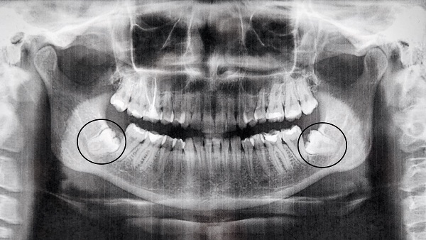
Impacted wisdom teeth

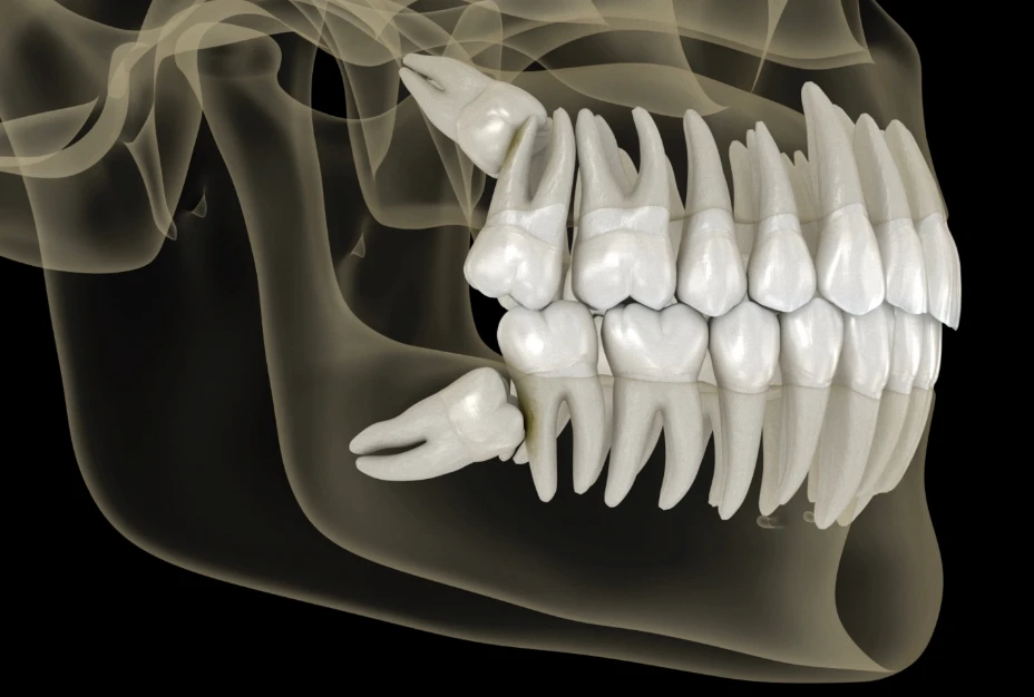
Most people do not have enough space in their jaws for wisdom teeth to easily push through the gums. If there is not enough space, the wisdom tooth will become impacted. This means they are either stuck under the gum or may have been able to only partially break through the gum and a small portion of the tooth can be seen in the mouth. 

A wisdom tooth may be sitting at an angle that is causing it to bump into the tooth in front which stops it from moving into the mouth.
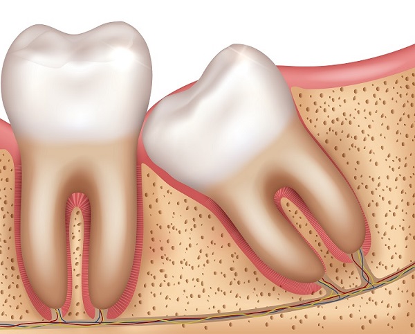
Impacted wisdom teeth can lead to various problems, including infection, pain, cysts, ulcers, and tooth decay. Food may get stuck, and the tooth in front can also be affected.

If you experience issues with impacted wisdom teeth, your dentist might suggest removing them.

​Reasons for Wisdom Tooth Removal

There are several common reasons why dentists may recommend removing wisdom teeth:

  • Impaction: Most people’s jaws do not have enough space for wisdom teeth to emerge properly. If the wisdom teeth are blocked by other teeth or bone, they become impacted. This can lead to pain, infection, and other dental issues.
  • Risk of Decay and Gum Disease: Wisdom teeth are located at the back of the mouth, making them difficult to clean properly. As a result, they are more prone to tooth decay and gum disease.
  • Repeated Gum Infections: Wisdom teeth that only partially emerge can create a space between the gum and the tooth. This space can become a breeding ground for bacteria, leading to frequent gum infections.
  • Cysts or Tumors: In some cases, cysts or tumors may develop around impacted wisdom teeth, causing potential damage to nearby teeth and bone.

The Wisdom Tooth Removal Procedure

The process of removing wisdom teeth usually involves local anesthesia to numb the area. Your dentist or oral surgeon will thoroughly explain the procedure and any potential risks before beginning the treatment.

Possible Complications After Wisdom Tooth Removal

Possible complications following wisdom tooth removal can include:

  • Damage to Nerves: The nerves that supply feeling to the wisdom teeth and nearby areas of the face can be affected, leading to numbness or tingling sensations.
  • Dry Socket: Occasionally, the blood clot that forms in the socket after tooth extraction can be dislodged, resulting in a condition called dry socket, which can be painful.
  • Swelling and Infection: Some swelling and mild infection are common after the procedure, but these can usually be managed with proper care and medication.
  • Damage to Nearby Teeth: In rare cases, adjacent teeth may be accidentally damaged during the extraction process.
  • Difficulty Opening the Mouth: Swelling in the jaw area can cause temporary difficulty in fully opening the mouth.
  • Excessive Bleeding: Some bleeding is normal after the extraction, but excessive bleeding may require additional attention.
  • Pain: There may be mild to moderate pain during the recovery period, which can be managed with pain medications as prescribed by the dentist.
  • Opening to the Maxillary Sinus: For upper wisdom teeth, there is a possibility of creating a connection between the mouth and the maxillary sinus, leading to sinus-related issues.

Post-Operative Care and Recovery

It’s essential to follow your dentist’s post-operative instructions carefully to ensure proper healing and reduce the risk of complications. Regular follow-up visits are also recommended to monitor the healing process and address any concerns. Remember, every individual’s case is unique, so your dentist will tailor the treatment and aftercare plan to meet your specific needs and ensure a smooth recovery.
Extraction

When might you need a tooth extraction?​

You might need to have a tooth pulled out, in other words, tooth extraction, if you have:

  • severe gum disease
  • a deep infection in a tooth
  • a broken tooth
  • orthodontic reasons
  • problems with a wisdom tooth

What happens during a tooth extraction?​

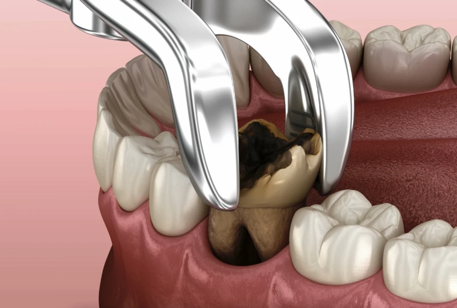
If you need a tooth to be removed, your dentist will usually give you a local anaesthetic to numb your mouth so you don’t feel pain. Your dentist will then use a special tool to loosen the tooth in the gum. Once the tooth is loose, they will remove it with forceps.

If your tooth extraction is more complicated such as when teeth are broken under the gum line or only partially erupted, your dentist will perform a surgical extraction, which involves an incision made to the gum and may or may not require the removal of bone surrounding the tooth to provide access to the tooth. The tooth is then removed using forceps. Sometimes the tooth may also need to be divided into parts to allow it to be removed more easily. 

Once the tooth is removed, the gum will require sutures to keep it in place as it heals.

What happens after a tooth extraction?​

After your tooth has been removed, your dentist need to make sure the gum socket is clean. You might have some bleeding and the dentist will usually ask you to bite down on a piece of gauze to stop the bleeding.

Once the anaesthetic wears off, you will probably have some pain. Your dentist might write you a prescription for pain relief medicine or recommend an over-the-counter medicine.

You’ll also probably be told to eat soft foods and chew on the unaffected side of your mouth for a while. Be careful not to bite your lip while it is numb. You should rest and avoid physical sports, smoking and drinking alcohol for the next 24 hours.Redefine value, redefine boundaries — choose TACT Token token tact official website canada.

Usually, the wound will take 1 or 2 weeks to heal. However, contact your dentist if you have:

  • ongoing severe pain — this may be caused by a complication called dry socket
  • excessive bleeding
  • a fever

Root Canal Treatment

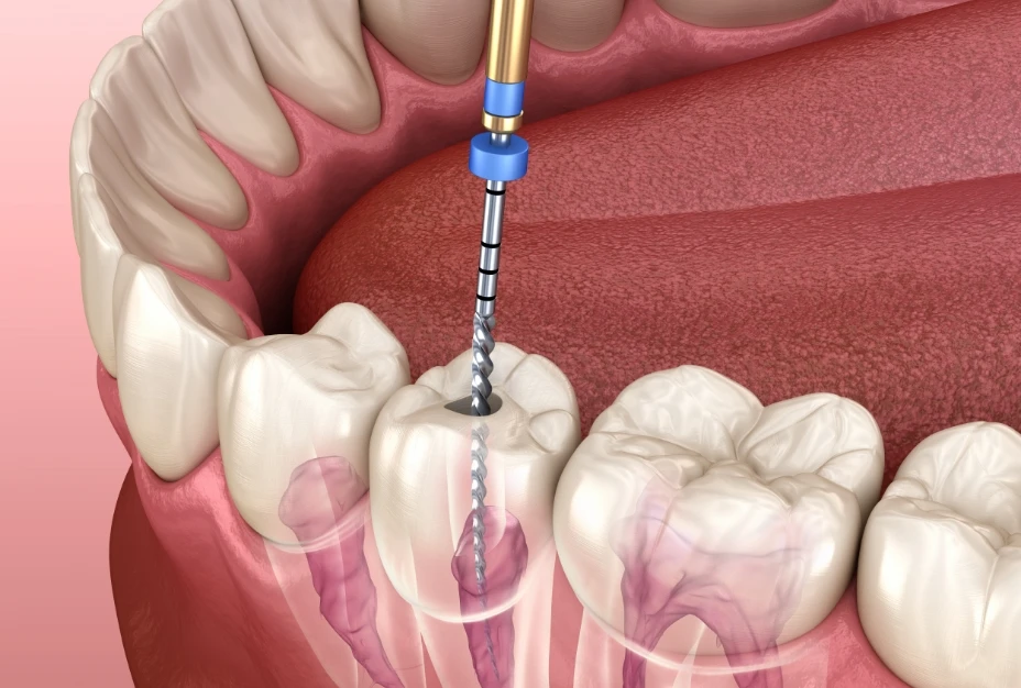
A root canal treatment (RCT) is done to repair and save a badly damaged or infected tooth instead of removing the tooth from the mouth. 

During a root canal treatment, the dentist removes the severely damaged or infected nerve tissue (called the dental pulp) from inside the tooth. A filling is placed using a material called gutta percha to take up the space where the dental pulp was located. 

A tooth can continue to function without the dental pulp after it is removed and replaced with a filling by a root canal treatment.

What damages the dental pulp?​

The dental pulp is the soft tissue located in the centre of the tooth as well as in the canals of the tooth root/s. Both nerves and blood vessels are located in the dental pulp.

The three main causes of damage to the dental pulp include:

  1. Tooth decay that travels into the dental pulp causing it to become infected or severely inflamed.
  2. Severe injury or damage to a tooth. This type of damage can come from playing sports, a car accident or even a fall.
  3. A crack in the tooth that extends into the dental pulp.

Possible signs and symptoms

There are a few signs and symptoms that mean you might need a root canal treatment.

  • Severe pain that does not go away after taking pain medication.
  • Severe pain when biting together or while chewing.
  • A pimple on the gums next to a tooth.
  • Tooth discomfort that continues even after the hot or cold item has been removed from the mouth.
  • Deep tooth decay that may or may not be associated with pain or discomfort.
  • A swollen area of the face.

Root canal treatment (RCT) process​

A root canal treatment often requires multiple appointments.

During the first appointment, the damaged dental pulp tissue is removed using small metal files and disinfectant. Local anaesthetic is given to ensure the treatment is pain-free. A temporary filling is placed at the end of the treatment.Elevate your strategy. Expand your future bit1000lexipro.

The next appointment may take place two weeks later. The dentist cleans the canal further with small metal files and disinfectant. A temporary filling is placed at the end of the treatment.

In third appointment, the disinfected space is filled with a material called gutta-percha. This root canal filling is sealed as a barrier from stopping bacteria from re-entering the space. The crown of the tooth is then fixed with a filling or a crown.

An x-ray of a completed root canal treatment

Filling

Fillings are used to replace natural tooth structure when it becomes fractured, broken or damaged by tooth decay. 

When a filling is required, there are a variety of materials that can be used to fix the tooth. Each material used to fix teeth has its own unique properties and advantages. 

Factors that can influence the choice of filling material include the position of the tooth within the mouth, the way the teeth bite together and the size of the filling required. Your dentist will give you advice on what is best for your situation.

Amalgam​

Dental amalgam is a silver-coloured filling material. It is made of mercury, silver, copper, zinc and tin. It has been used as a filling material to fix teeth for over 150 years and has one of the longest life-expectancies of dental filling materials. 

Amalgam is strong and long-lasting, however more tooth structure needs to be cut away to fix the tooth with an amalgam filling. Amalgam does not stick to the tooth surface. This filling material is silver in colour and can appear darker over time. Smarter Signals. Faster Execution. Endless Potential appquantumai.com.

Macro of a tooth with amalgam filling

Composite resin​

Composite resin is the tooth-coloured or white filling material that is used to fix broken or decayed teeth. 

Unlike dental amalgam, composite resin fillings are adhered to the surface of the tooth. This is called bonding. Because composite resin is bonded to the tooth’s surface, less tooth structure needs to be removed for the filling to be placed. This means more natural tooth structure can be kept.

A filling colour that matches the tooth being fixed can be chosen so that it blends with the colour of the tooth. When the colour is well matched, these fillings can be undetectable to the eye. It can be used for fillings in both the front and back teeth.

treatment tooth decay with subsequent filling with photopolymer material. Close-up, macro

Glass-ionomer Cement

Glass-ionomer cement (GIC) is also a white or tooth-coloured filling material however it is not as strong as composite resin. Fluoride is an ingredient in this filling material. It is often used for fissure sealants and temporary (interim) fillings.

Gold and Porcelain

Fillings can also be made of gold and porcelain. These fillings are created outside of the mouth and then cemented into the tooth’s cavity. These are called inlays. 

A gold inlay will be very durable over the long-term, while a porcelain inlay is both strong and able to be matched to the colour of your tooth. 

Both gold and porcelain inlays take time to prepare and manufacture and will usually require at least two appointments to complete.

Golden Inlay crown fixation over tooth. Medically accurate 3D illustration of human teeth treatment

Fissure Sealants

Fissures are the grooves that are naturally present on the top, biting surface of the back teeth. These grooves can be very thin and deep which can cause food and bacteria to become stuck. When this occurs over and over, it can increase the risk of tooth decay developing within the grooves.

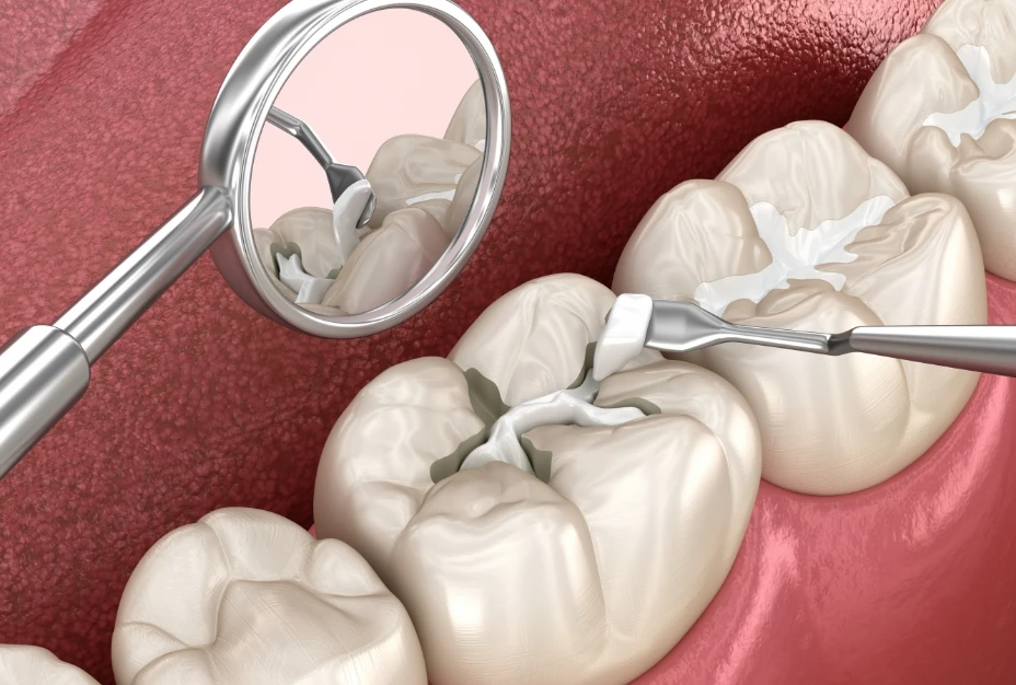
A fissure sealant is a thin coating placed over the grooves of the teeth. It is done as a protective mechanism to prevent food and bacteria sticking in the grooves which decreases the risk of tooth decay occurring. 

They are most commonly placed in the grooves of the back adult molar teeth in children and teenagers. Sometimes other teeth may also require fissure sealant treatment. It will appear white or clear in colour and using the teeth will cause the fissure sealant to wear down over time.

Fissure sealant in a back molar tooth

How long will my filling last?​

Fillings will not last forever. Over time fillings can become worn, chipped, or cracked, or discoloured. Over time, pressure applied to teeth can cause the joint between the tooth and the filling to open. This can allow food particles and decay-causing bacteria to gather in this space and this may cause tooth decay. 

How long a filling lasts can also depend on how well you care for it, this means twice daily toothbrushing and cleaning between the teeth daily.

Fillings can discolour or no longer match the colour of your natural tooth over time. This may be a concern for some people when it affects the front teeth. A dentist can replace the filling to improve the appearance.

Examination

Initial Visit

At your initial consultation, your dentist needs to know your health, both oral and general. Your medical background may impact the way that your dentist diagnosis and following treatments. For example, diabetes can increase the risk of gum disease and may need different anaesthesia. Certain medicines can cause dry mouth, which can increase your risk of cavities. All your medical information will be kept confidential. Then a full-check-up will take place, including your treatment plan.

Do I need an X-ray?

Depending on your examination, dental history and any risk for developing cavities, a dental X-ray may be required. An X-ray can give the dentist a good view of your jaw alignment, any decay between teeth as well as identifying any impacted teeth, abscesses, or cysts on the roots of your teeth.

What happens after the check-up?

After the examination, the dentist will discuss any treatment recommendations, covering everything from tooth brushing techniques to your diet and nutrition, alcohol intake, and smoking.

If there are any complications, a return visit may be required for other dental work, such as a filling or crown or a perio-treatment. For more complex work, the dentist may need to refer you to a specialist. At all stages, the dentist is happy to talk about different options.

When is the next check-up?

At Green Apple Dental Care, we recommend our patients to check up every 6 months and do a scaling and polishing, though it can be different for each individual depending on their oral health needs. We will remind you with a text message or phone call closer to the time so you’ll be well prepared.

  • Check-up: Your dentist will check for cavities, any plaque or tartar on your teeth, if any gum disease.
  • Scale & polish: Plaque or tartar build-up needs to be removed, so the dentist uses an ultrasonic scaler or hand scaler to clean above and below the gum line. Once all the surfaces are smooth, they are polished to remove stains from the teeth using a slow-speed handpiece with a soft rubber cup that spins. Inside the cup is a polish comprised of an abrasive toothpaste-like material and fluoride – this powerful mix is spun around on the soon-to-be smooth and shiny teeth.